Imaging endocytic vesicle formation at high spatial and temporal resolutions with the pulsed-pH protocol
Nature Protocols (2020)
Silvia Sposini, Morgane Rosendale, Léa Claverie, TN Ngoc Van, Damien Jullié & David Perrais
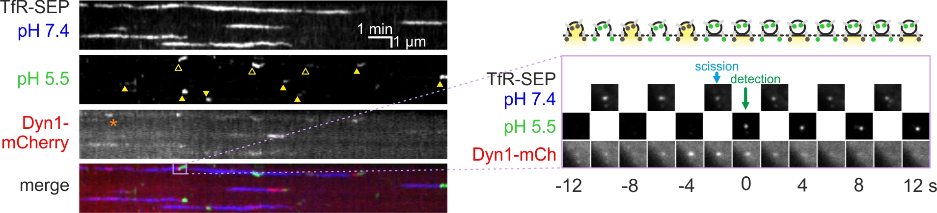
Endocytosis is a fundamental process occurring in all eukaryotic cells. Live cell imaging of endocytosis has helped to decipher many of its mechanisms and regulations. With the pulsed-pH (ppH) protocol, one can detect the formation of individual endocytic vesicles (EVs) with an unmatched temporal resolution of 2 s. The ppH protocol makes use of cargo protein (e.g., the transferrin receptor) coupled to a pH-sensitive fluorescent protein, such as superecliptic pHluorin (SEP), which is brightly fluorescent at pH 7.4 but not fluorescent at pH <6.0. If the SEP moiety is at the surface, its fluorescence will decrease when cells are exposed to a low pH (5.5) buffer. If the SEP moiety has been internalized, SEP will remain fluorescent even during application of the low pH buffer. Fast perfusion enables the complete exchange of low and high pH extracellular solutions every 2 s, defining the temporal resolution of the technique. Unlike other imaging-based endocytosis assays, the ppH protocol detects EVs without a priori hypotheses on the dynamics of vesicle formation. Here, we explain how the ppH protocol quantifies the endocytic activity of living cells and the recruitment of associated proteins in real time. We provide a step-by-step procedure for expression of the reporter proteins with transient transfection, live cell image acquisition with synchronized pH changes and automated analysis. The whole protocol can be performed in 2 d to provide quantitative information on the endocytic process being studied.